
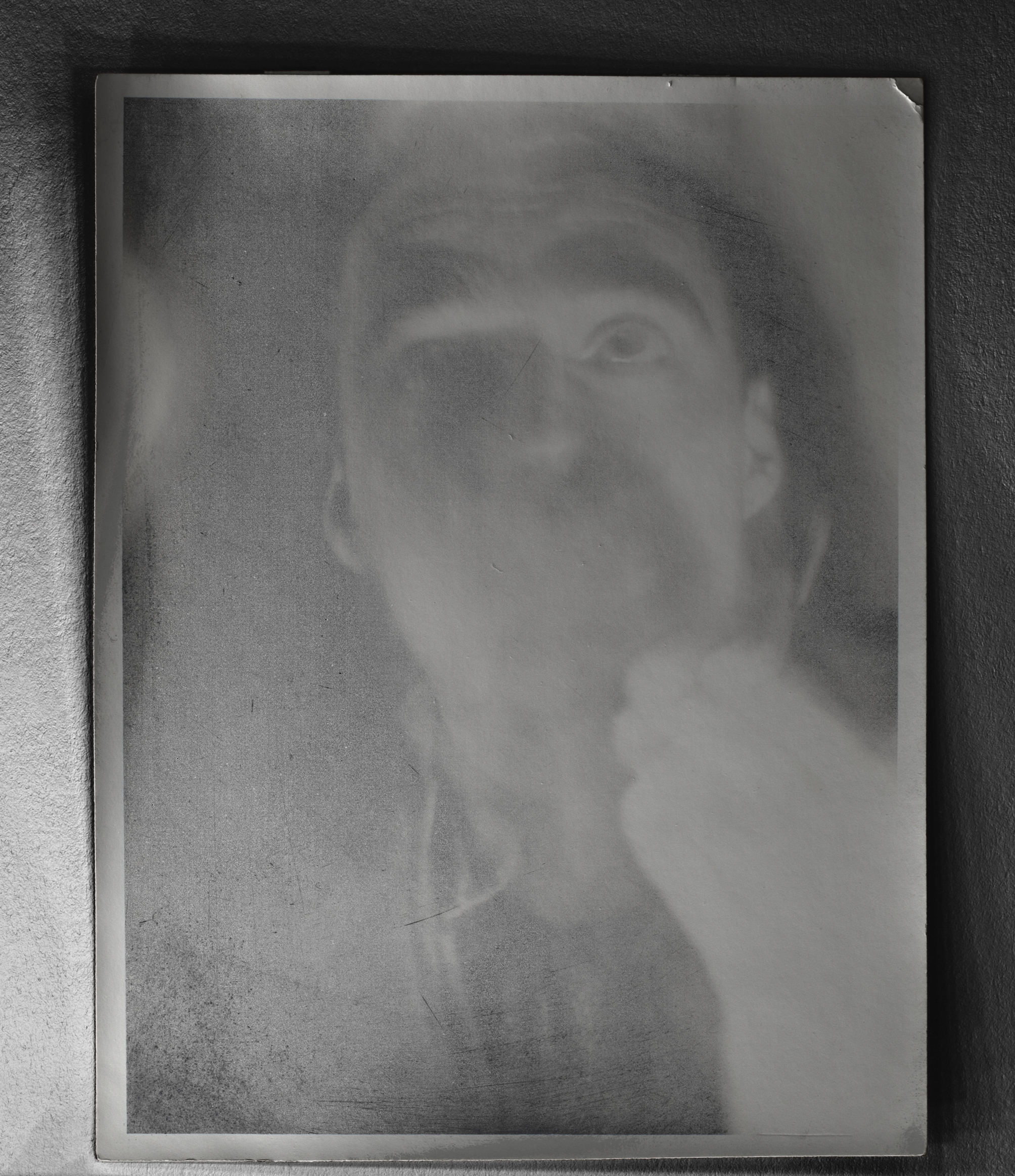
Front / Recto
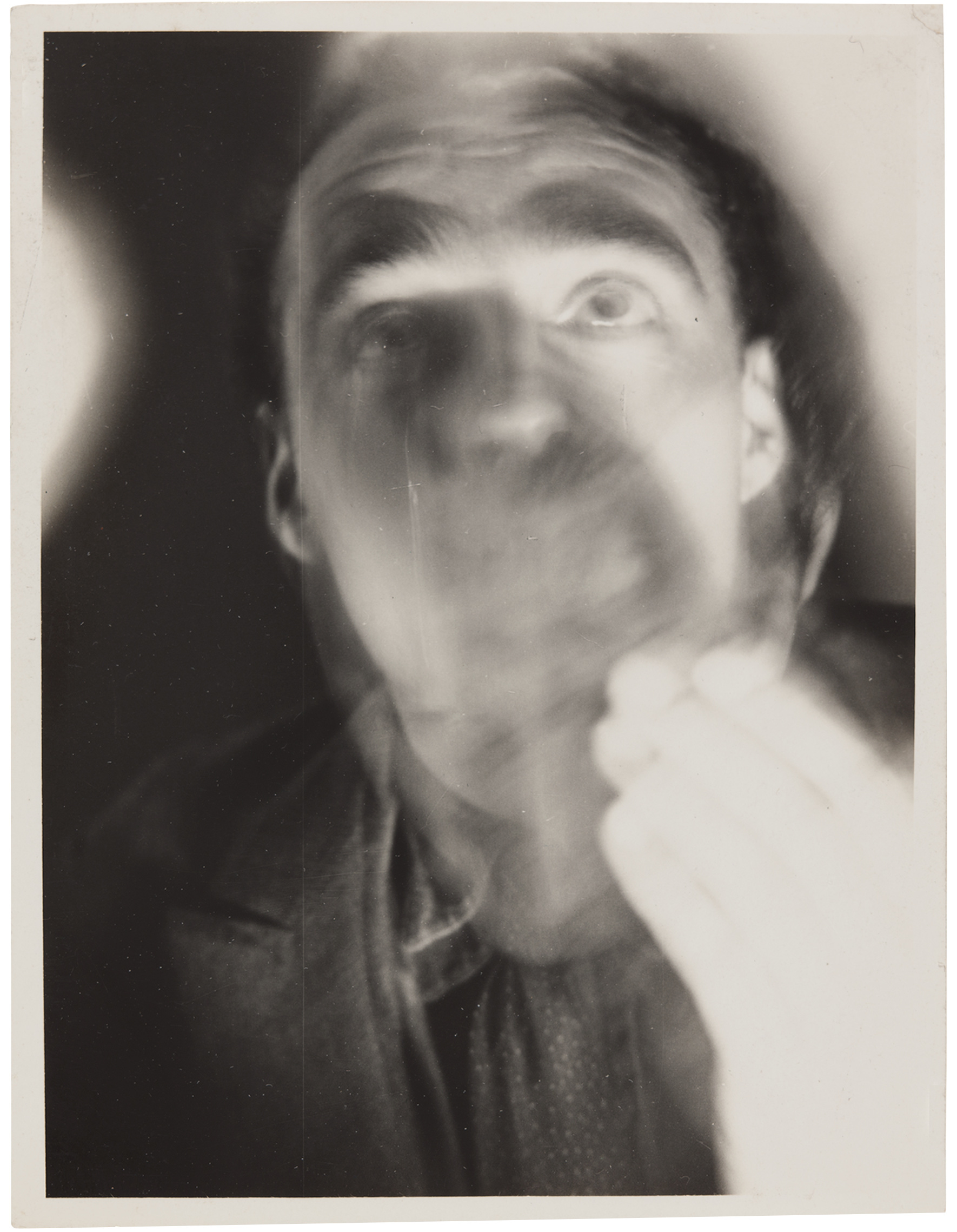
- Title Portrait of a Man Smoking
- Negative Date 1931
- Print Date 1931–39
- Medium Gelatin silver print
- Dimensions Image 4 3/16 x 3 1/8" (10.7 x 8 cm)Sheet 4 7/16 × 3 3/8" (11.2 × 8.6 cm)
- Place Taken Dessau
- Credit Line Thomas Walther Collection. Gift of Thomas Walther
- MoMA Accession Number 1917.2001
- Copyright © 2015 Makoto Yamawaki
Back / Verso
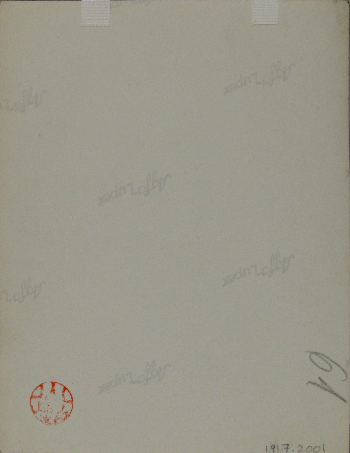
- Mount Type No mount
- Marks and Inscriptions Stamped in red ink on sheet verso, bottom left: Yamawaki. Inscribed in pencil on sheet verso, bottom right: 61.
-
Provenance
The artist; to Tom Jacobson, San Diego, 1985 [1]; to Howard Greenberg Gallery, New York, 1996 [2]; purchased by Thomas Walther, September 24, 1996 [3]; given to The Museum of Modern Art, New York, 2001.
[1] MacGill/Walther 2001(4), p. 15; and Alicia Colen (Howard Greenberg Gallery), e-mail to Maria Morris Hambourg, October 25, 2013.
[2] MacGill/Walther 2001(4), p. 15; and Colen, e-mail to Hambourg. The gallery acquired the print for a Yamawaki exhibition.
[3] MacGill/Walther 2001(4), p. 15; and Howard Greenberg Gallery invoice no. 96-0624, September 24, 1996.
Back Printing
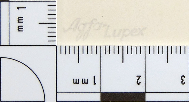
Detail showing Agfa-Lupex manufacturer logo printed in ink on the verso of the photograph. In image processing, contrast was adjusted to enhance the readability of the logo. The area of detail is 1 x 3 cm. Department of Conservation, MoMA
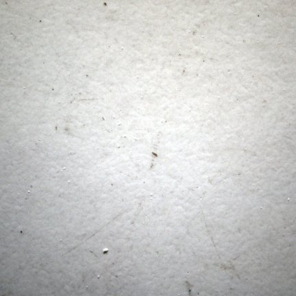
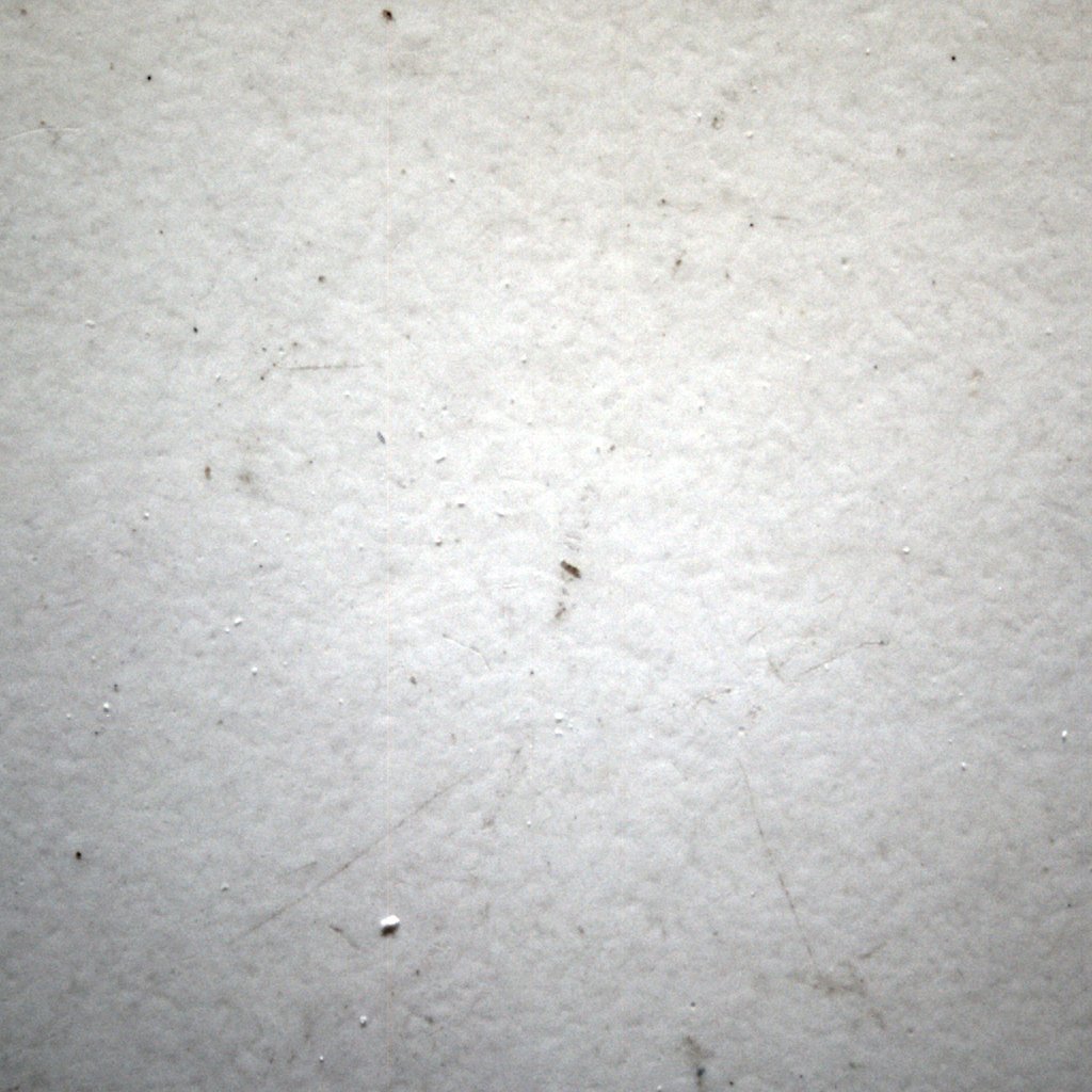
Surface
- Surface Sheen Glossy
- Techniques Retouching (additive) Contact print Retouching in negative
-
PTM
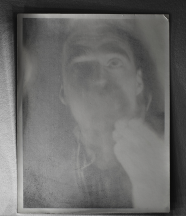 View of the recto of the artwork made using reflectance transformation imaging (RTI) software, which exaggerates subtle surface details and renders the features of the artwork plainly visible. Department of Conservation, MoMA
 -
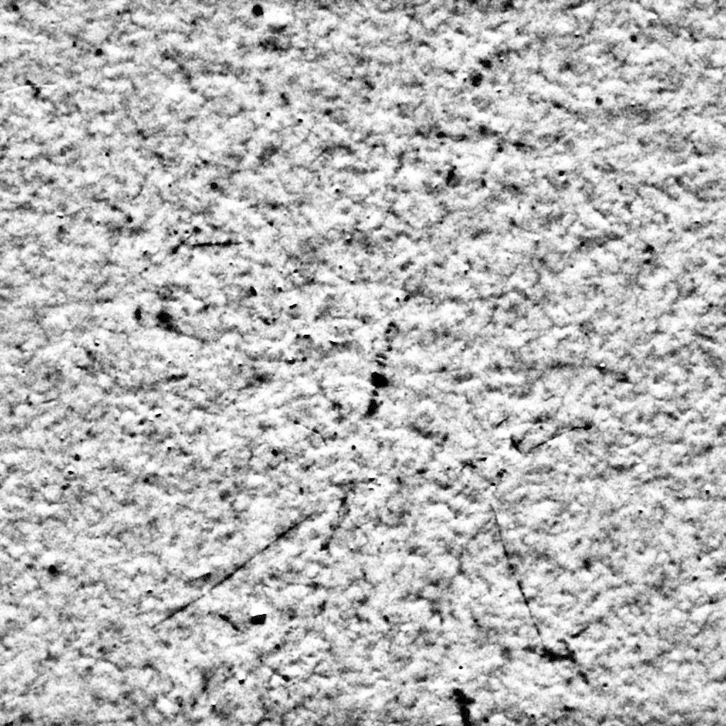
Micro-raking
 Raking-light close-up image, as shot. Area of detail is 6.7 x 6.7 mm. Department of Conservation, MoMA
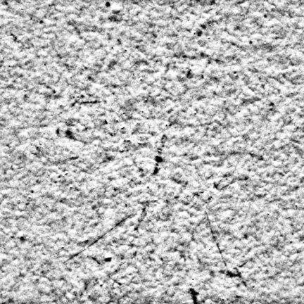 Raking-light close-up image, processed. Processing included removal of color, equalization of the histogram, and sharpening, all designed to enhance visual comparison. Department of Conservation, MoMA
Paper Material
- Format Metric
- Weight Single weight
- Thickness (mm) 0.17
- UV Fluorescence Recto negative Verso negative
- Fiber Analysis Softwood bleached sulfite 87% Hardwood bleached sulfite 1% Rag 7% Bast 4%
- Material Techniques Developing-out paper Back printing Gaslight paper
-
XRF
This work was determined to be a gelatin silver print via X-ray fluorescence (XRF) spectrometry.
The following elements have been positively identified in the work, through XRF readings taken from its recto and verso (or from the mount, where the verso was not accessible):
- Recto: P, S, Cl, Ca, Zn, Sr, Ag, Ba
- Verso: Al, Si, P, S, K, Ca, Fe, Zn, Sr, Ba
The graphs below show XRF spectra for three areas on the print: two of the recto—from areas of maximum and minimum image density (Dmax and Dmin)—and one of the verso or mount. The background spectrum represents the contribution of the XRF instrument itself. The first graph shows elements identified through the presence of their characteristic peaks in the lower energy range (0 to 8 keV). The second graph shows elements identified through the presence of their characteristic peaks in the higher energy range (8 to 40 keV).
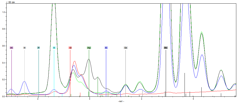 Areas examined: Recto (Dmax: black; Dmin: green), Verso or Mount (blue), Background (red)
Elements identified: Al, Si, P, S, Cl, K, Ca, Ag, Ba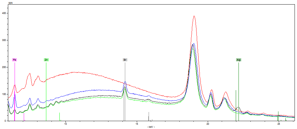 Areas examined: Recto (Dmax: black; Dmin: green), Verso or Mount (blue), Background (red)
Elements identified: Fe, Zn, Sr, Ag
In Context
Related People
-
Artist